 AttendMe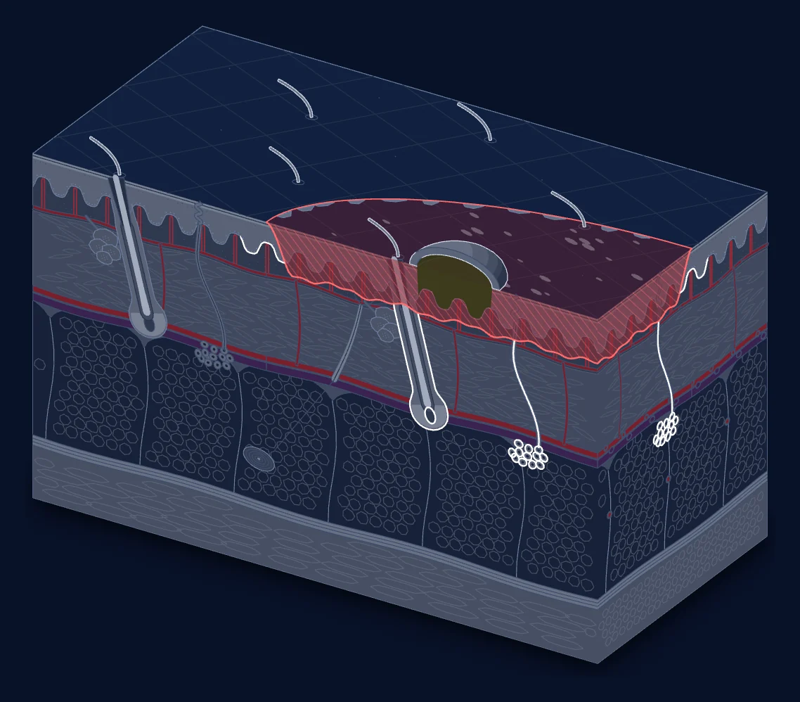
Learn from the underlying structure
The figure explores skin layers and a superficial dermal burn. It is a still image, not an interactive diagnostic tool.
Inspect the visual study
Use this still image to review the intended teaching design. The useful output is a question about the labels, boundaries or source detail a reviewed version must explain.
Look at the visual structure
Inspect the surfaces, layers and highlighted region in the illustration. This is a still image. It has no controls for assessing a wound or changing a physiological model.
Compare with an anatomy source
Open the NCI anatomy illustration linked below. Compare the intended layer structure with that reference. Record a boundary or label that needs revision rather than inferring a clinical classification from the development image.
Define the review task
A useful review note reads: 'Visual element to check: [identify it]. Source detail: [locate it]. Label or boundary to revise: [describe it]. Clinical-review status: pending.' The teaching release needs explicit source provenance, revision and independent review.
Source and review
The source anatomy references are the NCI skin illustration and Gray's Anatomy (1918). This adaptation remains a development study. Revision and independent clinical review must precede a reviewed teaching release.